
Processing
Likes
Comments
Share


@Masshysteria
Follow
Day 62 Update: Water and food. Pics uploaded from days 58 & 62. Daily journal to be updated soon along with photo details.
Days 63-69 Update: Nothing to do really but keep an eye on water and when to harvest. Letting the smaller three use up their stored energy rather than give more food. The big guy is still going to get nutrients because there’s a while left. Sometime this coming week I’ll chop down the shorter three plants.
Trichomes are about 80% cloudy, but very little amber.
Day 70 Update: nothing new to report.

Likes
8
Share


@Hiroots420
Follow
28/04/2021 (Day 43)
Se realizo un nuevo cambio de solución, las plantas ya están tirando pelos y los leds están al 80%, se observo un gran crecimiento radicular y las paredes del tallo son increíblemente gruesas y duras.
03/05/2021 (Day 48)
Las plantas vienen con un crecimiento optimo pero la flora se está tardando demasiado en hacerse presente, se estima que es debido al estrés que tuvo en semana 3 de vege produjo un retraso de algunos días.
Esta semana se incorpora Safe Roots al sistema con la finalidad de que deje la solución lo mas limpia posible.
También se realizaron bastantes defoliaciones ya que no existía penetracion de luz teniendo en cuenta el tamaño que están ocupando (85% del espacio de cultivo a 30/35 cm de altura)
05/05/2021 (Day 50)
Se realizo un nuevo cambio de solución, esta vez utilizando safe roots para limpiar todo el sistema hidroponico.
600w de Citizen a 30cm en 160x80 y la plantas parecieran que si les doy mas luz van a usarla, es increíble la tolerancia a la alta intensidad de ppfd que tienen, gracias a esto logramos subsanar esa semana de raíces marrones.
La temperatura bajo drásticamente y nos dejó el indoor en unos 22 grados, donde todo marcha perfecto.


Likes
3
Share

@SMOKINGMONKEY
Follow
WEEK 4
Day 29 Minor defoliation six large fan leave taken from
each plant now 19 inches
Day 30
32 oz water am Lazy susan remove and plants placed on
milk crates 550 PAR 6X Light
Day 32
32 oz water am
Day 33
6 large fan leaves taken from each plant. 21-23 inches
Day 34
40 oz water am

Likes
Comments
Share


@jojopfoh
Follow
They are really starting to fill out. I am seeing great results after every water and every feed. I will be transplanting them into 2 gallon pots in the next 2 weeks.

Likes
13
Share

@sanibelisl
Follow
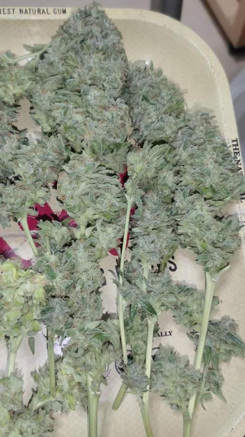
Mulberry F3 Day 84
Well this grow is coming to an end not much left to do here. Just waiting on some amber trichomes to show up and she will be finished. She has slowed way back on water consumption and the trikes are starting to stack up nicely on her flowers. Stopped nutrients two waterings ago and started molasses instead. Pretty sure my next update will be a harvest one. Thanks to everyone who followed this grow. I hope everyone is ready to go Bananas! Hahaha. Thinking my next grow will be a Purple Strawberry Popecicle.
Likes
1
Share


@Zugzug
Follow
just water no food
Here is a link to where you can find this
https://2fast4buds.com/us/seeds/gorilla-cookies-fast-flowering
Likes
15
Share


@DeepRootsGrowTrees
Follow
AUTO MOON ROCK / DIVINE SEEDS
WEEK #11 OVERALL
WEEK #6 FLOWER
This week she started to stack buds are getting dense and covered in trichomes, she's smelling great!! Stay Growing!!
Thank you for stopping by and taking a look it's much appreciated!!
Thank you DIVINE SEEDS!!
AUTO MOON ROCK / DIVINE SEEDS
Likes
52
Share


@SlowpokeFuegobud
Follow
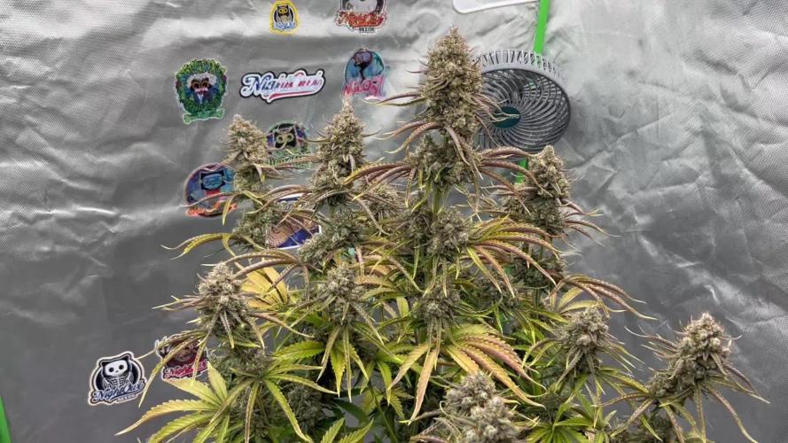
Cream Brulee Auto was my pleasure to grow, I definitely will grow this one again! The weed is nearly finished by the time I write this, one of the strains which is gone very fast.. 😍
I didn't have to do any LST nor much defoliation, next time I would feed her a bit more, but that's was pretty much a smooth grow with the Cream Brulee girl! 🍮
She grew all by her own, to be a beautiful queen! 👑
Thank you, Hypno Seeds, this is amazing work!! 🙏 💚
Thank you, GD community! 💚💚💚 It's an honor to grow together with you!! 💚💚💚💚💚💚💚

Likes
Comments
Share


Likes
17
Share


@RDWCGrowing
Follow
Week 1 Day 1 - 8/12/2023
1st Water change Day! Such a special time it is when you remove the little bit of Nutes that you gave them as an appetizer and you give them their first real meal.
Added
39 Gallons of Water to my system
SILICA= .5mil/Gal = 19.5 = 20mil
Root Drip = 1mil/Gal = 39mil
Cal Mag= .25mil/Gal = 9.75 = 10mil
FLoraMicro= 3.0mil/Gal = 114mil
FloraGro = 2.0mil/Gal = 78mil
FloraBloom = 2.0mil/Gal =78mil
ORCA= .5mil/Gal = 19.5 = 20mil
Week 1 Day 2 - 8/13/2023
Everything is looking good the roots are making their way to the water and the new grow is looking nice and green.
Week 1 Day 3- 8/14/2023
Everything is right on track, they are looking beautiful and in the praying position all leaves happily lifting towards the light.
Week 1 Day 4- 8/15/2023
A little worried today her birth Twin the BA I am growing out is looking great and is raised towards the light and this one is just slightly under.. Will keep an eye on Her.
Week 1 Day 5- 8/16/2023
Walked in and the humidity was under 60.... ohh noooooo.. So I added 2 humidifiers to the tent and attached them to my InkBird controller which is set to 62.
Also looking at the roots and she has some poking out the bottom but just not in the water yet.. Luckily we are set for 14 days before next water change so the system will stay stable and her sister already has roots in the water so she should only be a day or two behind.. we will just keep tracking but she is delayed.
Week 1 Day 6- 8/17/2023
Roots in the Water!!! Huston, we have a successful launch. This grow is on!
Humidity was a little low this morning so I refilled the humidifiers. Other than that the temp looks great, the PH looks good, the PPM looks good the plant is in the praying position and all damage from the little drowning from over filling the cloning machine seems to have been fixed. Happy Happy.
Week 1 Day 7- 8/18/2023
Yay.. week 1 in the books, roots in the water growth has started. Everything for growth and environment is looking good and on track, there are a couple of mutations with this Lady will keep an eye on those leaves.
IMO this grow is going A lot during this week 1 then week 1 of the last grow when I had them drowning.
Really excited on how this grow is going to come out.


Likes
25
Share


@eldruida_lamota
Follow
Ya estoy de nuevo familia, vuelvo actualizar, la primera semana de floración de estas Casey,s rollex O.G. Están gozando con un buen crecimiento y buena vegetación.
Ya marcó su sexo 3/3 hembras, las condiciones climáticas si están siendo óptimas en este espacio.
.
La humedad la tengo al 50% la temperatura está entre 21/25 grados , y el ph está en 5,8/6,0.
.
AgroBeta:
0,6 ml x L Flowering black line , vía radicular.
0,3 ml x L Tucán , vía radicular.
0,8 ml x L Génesis, vía radicular.
0,1 ml x L Betazyme, vía radicular.
0,05 ml x L Gold Joker, vía radicular.
0,2 ml x L Silver, vía radicular.
.
Hasta aquí es todo amigos, buenos humos fumetillas💨💨💨.


Likes
11
Share


@Gonach
Follow
Al principio de la semana se subió la luz a 70 cm aproz de la planta, reduciendo la potencia al 50%. Se regó a cada una de manera foliar con Alg-a-mic de biobizz para regularizar la deficencia de nitrógeno. A mitad de semana se regó con 3 ml x litro de biobizz a c/u. Cada planta consume aproximadamente 1.6 lt.
Al final de la semana se cambio el ciclo a 12/12 y los resultados como se puede ver en el video.
Gracias a todos x sus posteos....


Likes
2
Share

@in_a_few_weeks
Follow
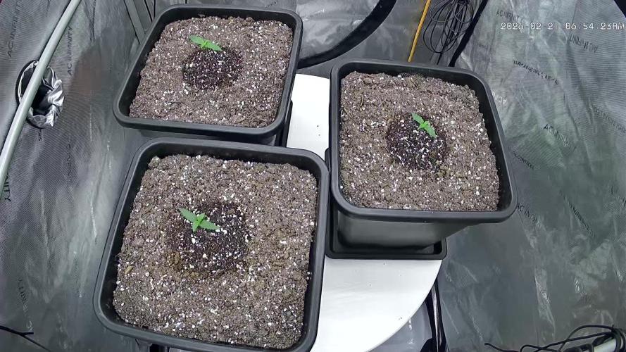
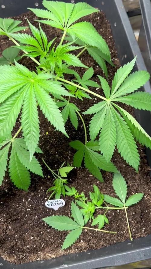
I seperated them now. The bigger one stays in the 90ltr Pot. The other got in a 13ltr pot. Later in 20ltr. Topping the small on in 1 or 2 weeks. Than LST and 12/12. Its a bit hot the next weeks. Have to look more after the plants.
Likes
3
Share


@CanarianGrow92
Follow
Week 5 already, thats why they call it fastbuds... In no time its harvest time! We had some bugs (trips) running around, i think i took them from outside as i was working on my garden, my outdoor plant is also affected, that happens for being a dirty old man 🤦🤷 but not much we can do now, i tried to clean the leaves a bit and remove some, but i think its better to harvest and clean the room as i dont eant any pesticides on the buds, but for the rest quite ok, we gave aome extra pk and bloom this week so lets see how the plants do next weeks 😁
Processing
Likes
11
Share


Processing
Likes
22
Share


@Heavier
Follow
Week 11: Wow such a busy week i'm sorry i am late on this one. Loads of humidity with all the new growth there is so much more plant transpiring and its has really drove my humidity up. So what i am doing is pruning stems and leafs that i don't think will mature in time and this should also help to limit the humidity i will have once things start to set heavily I want to harvest and not have to fight mold. But this aggressive pruning i think might be limiting my harvest. I might have to invest in a dehumidifier. Pictures will be updated and added later. With all the mulching i've done its really helped lessen my work load as i do not have to water as often.
Not much to write about here. Life has been busy and the plants are just stretching and growing every day. I have raised my lights so often these past few weeks.
