


Likes
Comments
Share


@Zeravlab
Follow
1/8/24- Beg of week - hairs are beginning to turn red, trichomes are mostly all clear or white. I think at least one more week. Cleaned the bucket and trimmed some.

Processing
Likes
29
Share


@Lennys_Laboratory
Follow
Feed Started With Tap Water Left Overnight For Chlorine/Chlorides To Evaporate.
Next Day.
Starting EC of our water is 0.10-0.15.
I added 1ML of Regenaroot Per Litre. Then Used Canna Terra Flores Until EC measured 0.7.
I then used the mighty bloom enhancer by the ml until my EC measured 1.3
Always Give A Little Sprinkle Of Great White Into The Mix
Finally i PH'd the mix to 6.2. Made sure the feed was of adequate temp (21c) and continued to feed.
😎

Likes
17
Share


@CanadianFillmore
Follow
Week 5 begins for Mandarin dreams and Divine storm. All four ladies look happy and healthy with minor deficiency signs. All four are bulking up and frosty as my windows (it's -21 °c)!
The Mars-Hydro TSW2000 is performing awesome, very happy with it. Thank you Kaoritracy for hooking me up!
Thanks for stopping by growfessors 👽🌳💚


Likes
2
Share
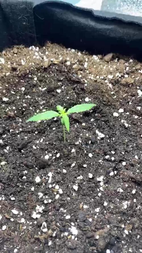

@Sativa1444
Follow
HAZIRLIK
Dolabı yapmak bir aydan fazla ileri sürdü; aydınlatma, havalandırması, iç kaplaması, montajı, elektrik kabloları, fişleri, soketleri, prizleri vb. Tüm bunların ardından, sonunda bitirmem ve kotiledonların görünenlerin görünmediğini gördüm. Burada yaşadığım mücadeleyi okuduğunuzda, heyecanımı ve mutluluğumu paylaştığınızı düşünüyorum… =)
//LED'LER//
Torch LED'leri kullanıyorum ve bu LED'ler fiziksel olarak oldukça büyük olduğu için, dolapta ciddi miktarda dikey alan kaplıyorlar. Toplam 70 cm'lik aralıklardan 50 cm'ye düşüyorlardı ve toprak birleştirmeyle yaklaşık 40 cm'ye düştü, bu da başlangıç büyümesi için neredeyse hiç yer bırakmadı. Bunu çözmek için Torch LED'lerini plastik yuvalarından çıkardım ve bunları ayrı ayrı boyutlandırılmış ahşap plakaya düz bir şekilde monte ettim. Bunu yaparak yaklaşık 15 cm'lik alandan tasarruf sağladınız. Ancak dolap yapısı ve Torch LED'lerinin kendisi nedeniyle ciddi ısınma sorunlarının belirtileri.
//KABİN//
LED'leri açtığımda, 26,5°C'lik iç havayı alıp 37–38°C'de dışarıya atıyorlardı. Her lamba bana yaklaşık +4°C'lik bir ısı artışı sağlıyordu.
Durum böyle dönemde fan kapsamını artırdım; bu sıcaklıkta yardımcı oldu, ancak daha sonra içerideki nem 35–30'a, hatta %25'e düştü.
Ne yeteneğimi düşünürken, 12V çift taraflı bir Peltier modül kullanmayı planladım: soğuklukta bakır borular sararak bunu fan destekli bir sıcaklığa dönüştürdüm. Aslında işe yarayabilirdi, ancak sıcak ortamdan ısıyı uzaklaştırmak için ihtiyacım olan sistem çok hantal hale geldi. O yerde, "Büyüme hayali bitti" dedim ve vazgeçtim.
Birkaç gün sonra "Neden olmasın?" diye düşündüm ve dolabı balkona taşımaya karar verdim. Sonuçta, gizliydi - kimse içinde ne olduğunu bilmeyecekti - ve balkon uydu yoluyla mükemmeldi.
Dolap balkonunda olacağından, iç kısım ısıdan, yağmurdan, bölgelerden ve diğer zararlılardan tamamen yalıtılması gerekiyordu. Tüm delikleri ve ortamları sıcak silikonla kapatırım. Daha sonra ısıl işlem köpüğü yerleştirdim ve her şeyi bölme filmle kapladım.
Tüm bunları yaparken termostat, ısıtıcı, nemlendirme ve vantilatör için hiçbir şey görünmemesi için tüm kablo bağlantılarını da gizledim. LED aydınlatma için tek tek veya hepsi birlikte kontrol edilebilen ayrı lamba yuvaları ekledim. Ayrıca dolap kapağı açıldığında tüm ana LED'lerin otomatik olarak söndüğü ve yalnızca küçük bir 5W LED'in açık kaldığı bir sistemim oluştu.
Vantilatörü dolabın sağ üst arka köşesine monte ederek egzozu diğer noktalara yönlendirdim. Vantilatörün gücünde, negatif basınç o kadar güçlüydü ki hava yalnızca girişten katılıyordu - başka hiçbir yerde 0,001 m³ bile hava kaçağı yoktu. (Bunu hava verileri için değil, zararlıları uzak tutmak için yaptım.)
//SICAKLIK VE NEM//
Dolabı balkona taşındıktan sonra bu sefer ciddi bir soğukluk sorunuyla karşılaştım. Geceleri sıcaklık 8-9°C'ye düşüyor, gündüzleri ise 24-27°C'ye kadar çıkıyor.
Elektrikli fanlı ısıtıcılar kullanmak istemedim çünkü havayı çok kurutuyorlar ve tehlikeliler.
Peltier fikrinden ilham alarak tekrar düşünürken çözümümü bir PTC ısıtıcı kullanarak buldum. Hava bağlantıları olan iki alüminyum bloktan oluşan bir PTC ısıtma elemanı yerleştirdim ve gelen tüm havayı bu kanallardan mülkler.
Sistemi 24,5°C'de açılıp 24,6°C'de kapanacak şekilde ayarladım. Bu şekilde sıcaklık sadece 1,5° tolerans içinde -24,5 ile 26,5°C arasında dalgalandı ve LED sayıları azaldıkça daha stabil hale geldi. PTC'yi termostata bağlayarak ısıtma sistemini tamamladım.
Gündüz sıcaklık sorunlarının çözülmesi için, elektrikler 11:45'te kapanıp 17:45'te tekrar açılacak şekilde programladım; böylece 6 saatlik bir kesinti süresini elde ettim. Bu süre boyunca kabin yaklaşık 24°C'lik dış havayı çekiyor, maksimum 27°C'ye ulaşıyor ve ardından tekrar soğumaya başlıyor; tam da bu noktada PTC devreye giriyor. Böylece
daha önceki büyük yangının sorunu tamamen çözülmüş olmuştu.
Ayrıca nemlendirmeyi termostata bağlayıp %70'e ayarlayarak nem sorunu da çözülürm.
Tüm bu süreci 1,5 ayımı aldı ama değdi.
1-2-3. güne kadar tek led 3. günde 10. güne kadar 3 ledide açtım ama bitkide büyüme problemi olduğunu gördüğüm için ışıkları söndürüp tek ışık 40cmden devam ettim.
Likes
5
Share


@AustinRon
Follow
TH 1Q2025 - Week 5 - Bolt 2
(ON Haze X Original Haze) X Northern Lights #2
“Todd’s Haze”
Objective
- 8 Female Plants, Topped ONCE @ Flip, 12” when topped
- Modified Sea of Green
Seeds Wet: 1139PM, 28.2.2025
Germinated: 2.3.2025
Flip: 21.3.2025
Harvest: 77 Days, DATE: 6.6.2025
Weeks Summary
- Continuing slow increase of EC & PPFD - Achieved EC: 1.8
- Allowing natural PPFD Increase (STRETCH), and providing modicum of intensity increase.
_________________________________________
__
Fri Apr 4, 2025
TH 1Q25 15:B:2:1
Light Intensity: [ 400, µMol/m2/s]
EC: [ 1.1, mS/cm]
Foliar: # 1 pt Spray Bottle @ Lights ON
- [x] CalMag Fuel: [ 2.5, ml]
- [x] Lush Green: [ 0.67, ml]
- [x] Kelpak (Auxins): [ 1, ml]
- [x] Photosynthesis Plus: [ 1.5, ml]
- [x] Quillaja 60 Powder: [scant]
- [x] Fertigation: 5:2:2:2:2:2:0
__
Sat Apr 5, 2025
TH 1Q25 16:B:2:2
EC: 1.3
- [x] EC: 1.3
- [x] Fertigation: [5.8:5.8:2.3:2.3:2.3:2.3:2.3] # [Primer A:Primer B:CalMag Fuel:Silica Skin:Lush Green:Root Anchor:Peak Bloom]
__
Sun Apr 6, 2025
TH 1Q25 17:B:2:3
- [x] EC: 1.4
Foliar: # 1 pt Spray Bottle @ Lights ON
- [x] CalMag Fuel: [ 2.5, ml]
- [x] Lush Green: [ 0.67, ml]
- [x] Kelpak (Auxins): [ 1, ml]
- [x] Photosynthesis Plus: [ 1.5, ml]
- [x] Quillaja 60 Powder: [scant]
- [x] Fertigation: [6.3:6.3:2.5:2.5:2.5:2.5:2.5] # [Primer A:Primer B:CalMag Fuel:Silica Skin:Lush Green:Root Anchor:Peak Bloom]

__
Mon Apr 7, 2025
TH 1Q25 18:B:2:4
EC: 1.5
Raised Dimmer to 77%
LightIntensity: [ 517, µMol/m2/s]
- [x] Fertigation: [6.3:6.3:2.5:2.5:2.5:2.5:2.5] # [Primer A:Primer B:CalMag Fuel:Silica Skin:Lush Green:Root Anchor:Peak Bloom]
__
Tue Apr 8, 2025
TH 1Q25 19:B:2:5
EC: 1.6
LightIntensity: [ 517, µMol/m2/s]
Foliar: # 1 pt Spray Bottle @ Lights ON
- [x] CalMag Fuel: [ 2.5, ml]
- [x] Lush Green: [ 0.67, ml]
- [x] Silica Skin: [ 2, ml]
- [x] Fertigation: [7.1:7.1:3.5:3.5:2.8:2.8:2.8] # [Primer A:Primer B:CalMag Fuel:Silica Skin:Lush Green:Root Anchor:Peak Bloom]
- [x] Terps Plus: [ 0.2, ml]
- [x] Photosynthesis Plus: [ 6, ml]
__
Wed Apr 9, 2025
TH 1Q25 20:B:2:6
Looking Relaxed, Happy, Lifted, Supple.
EC: 1.7
- [x] Fertigation: [7.1:7.1:3.5:3.5:2.8:2.8:2.8] # [Primer A:Primer B:CalMag Fuel:Silica Skin:Lush Green:Root Anchor:Peak Bloom]
- [x] Kelpak: [ 4, ml]
- [x] Photosynthesis Plus: [ 6, ml]
- [x] Terps Plus: [ 0.2, ml]
- [x] Quillaja 60 Powder: [scant]
__
Thu Apr 10, 2025
TH 1Q25 21:B:2:7
Light Distance: [ 23, in]
HOME Depot Run
- [x] Tube Insulation (Re-Fresh Chiller Insulation)
EC: 1.8 # MAX EC for GROW
Foliar: # 1 pt Spray Bottle @ Lights ON
- [x] CAL106 (Micronized Calcium Carbonate): [ 0.125, g]
- [x] Lush Green: [ 0.67, ml]
- [x] Kelpak (Auxins): [ 1, ml]
- [x] Photosynthesis Plus: [ 1.5, ml]
- [x] Quillaja 60 Powder: [scant]
- [x] Fertigation: [8:8:4.8:4.8:3.2:3.2:0.0:0.0:3.2] # [Primer A:Primer B:CalMag Fuel:Silica Skin:Lush Green:Root Anchor:Peak Bloom]
- [x] PCAL 1660: [ 0.5, gm] # Once or Twice/Week
- [x] Kelpak: [ 4, ml]
- [x] Photosynthesis Plus: [ 6, ml]
- [x] Terps Plus: [ 0.2, ml]
- [x] Quillaja 60 Powder: [scant]

Likes
4
Share


@letmegrow85
Follow
Und wieder ist eine Woche rum, 1ne von 3 habe ich geerntet, die anderen 2 lasse ich noch nen paar Tage stehen.
Soo, Nachtrag, die letzte Lemy85, da sind die Bud's richtig dick geworden.

Likes
85
Share


@Mr_und_Ms_Lue_Roots
Follow
Hallo zusammen,
wir stehen Anfang siebte Blütewoche und dieses wird für diesen Lauf auch die letzte sein, da wir erste Erfahrungen mit Budrot machen durften.
Allgemein waren die Daten die uns der Growhub angezeigt hat nicht alt zu verkehrt. Rückblickend war aber zu viel Masse und einige Bud´s zu tage gekommen und schlussendlich waren wir in der Feinabstimmung noch nicht im optimalen Bereich, ganz klar. Dennoch besteht auch bei uns noch Potenzial nach oben!
Gedanklich haben wir vieles vertiefen können und sind auch sehr froh über diese neue Erfahrung, auch wenn diese definitiv nicht positiv ist!
Zum Glück haben wir den Budrot Recht schnell ausfindig machen, großzügig Entfernen. Stetige Kontrollen haben wir geführt. Im Nachgang fing die Dame hinten Links durch Stress noch an zu HERM. Dahingehend haben wir im unteren Bereich für die letzten drei Tage auch unnützes herausgenommen. Auch zum Zeitpunkt der Feststellung des Budrot, haben wir jede Menge an Blattgut entfernt, um die R. Raumfeuchte auf mind. ca. 50 Prozent zu bekommen. Seit dem Feststellen, haben wir nur noch gespült mit Wasser.
Insgesamt sind wir dennoch froh, dass bis hierhin nicht alles im Abfall gelandet ist und zum Glück der Schaden nicht alt zu arg ist.
Negative Erfahrungen verbessern einen nur, wenn man natürlich nicht aufgibt. So etwas gehört leider auf dieser Ebene dazu, wir können ein Hacken dahinter machen.
Zudem hat es uns noch näher an unser recht aktuelles und erst benutztes Setup gebracht, was auch sehr viel Wert ist. 😉
So nun die Kulla Tränchen weggewischt und weiter geht es! Wir brauchen den Platz und kümmern uns erstmal darum xD
Revue gesehen, obwohl es an vielen Stellen nicht immer positiv war, ist unsere eigene Eischätzung zu diesem Tagebuch sehr positiv. Hier passierte viel und die Pflanzen sind doch auch wenn mit Umwegen an ihr Lebensziel gekommen. Wir steigen insgesamt eine Woche eher als angegebene mind. Blütezeit aus. Die Trichome sehen auch nicht schlecht aus, auch wenn wir sie selbst natürlich noch mind. Eine Woche noch gesehen hätten.
Der nächste Lauf wird besser, ganz sicher. Wir haben jetzt unser Zelt kennengelernt, die Anbauweise wird in Zukunft filigraner ausfallen ^^ Es war schon doch ganz gut gefüllt das Zelt xD
So nun das war es jetzt erstmal, wir wünschen allen Growmies ein mega entspanntes Wochenende und vielen Dank für euren Besuch!
VG

Likes
10
Share


@littlenugget
Follow
I checked the trichomes w. ~70X for the first time at the end of week 7 and they were clear. I checked again on the following day (day 51) and they had started turning milky so, I flushed with (with regular pH-ed water) until runoff was reading ≤150 ppm. I'm a little worried I'm late on the draw with flushing, but I'm seeing very few amber trichomes at this point. They are getting chunkier by the day! Let's hope I flushed well enough.. home stretch.
Likes
11
Share


@Bluemels
Follow
Tag 53: Ab heute beginnt die Blüte bei der Chocolate Haze. Obwohl ich sie vor 2 Tagen nochmals entlaubt habe, ist die Pflanze schon wieder enorm gewachsen 😳
Ich hoffe ich kriege sie in der Blüte gebändigt 😬
Likes
13
Share


@J_diaz420
Follow
Genética muy estable, floración rápida, hermoso color y bastante generosa 💚❤️

Likes
53
Share


@Mrs_Larimar
Follow
2025-04-21
Holy guercamole
these ladies keep getting fatter ( vertical)- and they dont look close to end at all
mindblowing
the room is filled with remaining 7 huge plants , and 2 photoperiods in vegetation for outdoor
everything runs smooth
i am refilling the tank every 4 days, and its so easy----
iam going to give them some booster in this week
xxxxxxxxxxxxxxxxxxxxxxxxxxxxxxxxxxxxxxxxxxxxxxxxxxxxxxxxxxxxxxxxxxxxxxxxxxxxxxxxxxxxxxxxxxxxxxxxxxxxxxxxx
Growing Environment Setup & Specifications
Lighting System
Model: Sanlight EVO 5-150
Quantity: Up to 5 units
Power Output: 320W per unit
Maximum Total Output: 1600W
Environmental Control
Management Unit: Growcontrol Growbase Pro
Humidity Management:
Dedicated Humidifier
Dedicated Dehumidifier
Temperature Control:
Primary: System-controlled
Secondary: Additional Radiator for supplemental heating
Air Management
Air Circulation: 2x Stand Fans
Air Extraction: Exhaust Fan with Filter System
Irrigation System
Primary: Autopot XXL Pot System
Water Storage: Flexi Tank
Root Zone Aeration:
Airdomes
Dedicated Air Pump
Growing Medium
Type: TERRA PRETA MJ-Mix
Nutrients
System: BIOTABS
Method: "Just Add Water" Strategy
Type: Organic-based feeding


Likes
1
Share


@GingerGarageGrow420
Follow
Day: 35 from sprout
Strain: Chill Out OG F4
Medium: Fox Farm Ocean Forest
Light: Vivosun VS2000 50%
Light Distance: 12 inches
Watering: By hand, ~12 oz daily
Nutrients: pH Perfect Advanced Nutrition Grow, Bloom, Micro 2 ml / L
Looking bushy. Did a little defoliation throughout the week


Likes
3
Share


@TS_Greens23
Follow
I have to say,,at the beginning of the Apricot Autos journey,,she had multiple tops,,and jumped out of the barriers like lightning,,,my sexy fingers,,managed to snap the top off what appeared to be the leader,,,and she instantly started flowering,,and didn’t stretch out like I thought she might have,,,,so to cut my losses,,I put her in the outdoor garden to finish up
Likes
9
Share


@Ukgrower99
Follow
Final week of flower she's only been given plane water she will be cut down in a few days the dark colour on the leafs are really showing
Likes
13
Share


@Drawer
Follow
Removed some buds that weren't getting any light, I ran into some issues regarding mold and that is most likely due to the fact that the RH % of my tent goes up to 70/80% at night.
I've removed all mold I could see (2 branches) and I am keeping a close eye on them, I've placed a new tower fan and another small adjustable fan in the tent for more airflow, my dehumidifier is coming in 2 weeks (around the time this grow is finished..)


